Cornea & Keratoconus
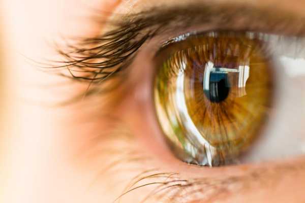
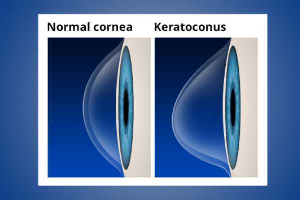
Let’s start by understanding what cornea is and what is its role when it comes to vision. A cornea is a clear, convex (dome-shaped) outer layer of eye that forms an important refractive surface where light rays enter and then focus on the retina. Furthermore, it also acts as a protective layer against dust, dirt, and germs.
There are many types of corneal problems that can affect your vision and cause problems with the cornea’s structure. Below are the two conditions that are most commonly seen in patients nowadays and effectively managed by the specialists at Centre for Sight.
Corneal Ulcer
A corneal ulcer is an open sore that occurs due to various reasons. Some reasons being infection, severe dryness of the eye, trauma, allergy, and incomplete eyelid closure. In all these cases, the bacteria or fungi enters cornea through minor erosions or abrasions and cause ulcer.
The occurrence of ulcer is more commonly seen in contact lens users. Common symptoms to look out for are decreased vision, redness of eyes, watering, swelling, pain and photophobia (extreme sensitivity to light).

Medical Management of Corneal Ulcer
If caused due to an infection, the first step of treatment is identification of microorganism causing this malady. A part of ulcer is mildly scraped for investigation. After identification, treatment targeted specifically at the causative agent is prescribed in form of eyedrops and many-a-times tablets.
Surgical Management of Corneal Ulcer
Uncontrolled corneal ulcer becomes a sight-threatening condition. In certain cases, surgical treatment is the only option left. If the ulcer is unresponsive to medical treatment or if the ulcer is large to begin with, a corneal transplant is done to save the eye. The purpose of corneal transplant here is to remove the microbiological load and prevent the infection from spreading to deeper structures of the eye.
Contact Us For Cornea & Keratoconus
To get in touch, complete this form and we'll get back to you as quickly as possible.
